A protruding bone at the base of your big toe represents one of the most common foot deformities affecting millions of people worldwide. This prominent bony bump, medically known as a bunion or hallux valgus, occurs when the complex biomechanical balance of your foot becomes disrupted. The condition develops gradually over time, causing the big toe joint to shift out of its natural alignment and creating a visible protrusion that can become increasingly painful and debilitating.
Understanding why this bone prominence occurs requires examining the intricate interplay between anatomical structure, genetic predisposition, biomechanical forces, and environmental factors. The metatarsophalangeal joint, where your big toe connects to your foot, bears significant weight during daily activities and walking. When this joint experiences prolonged stress or abnormal pressure patterns, compensatory changes begin to occur that ultimately result in the characteristic bone protrusion.
Hallux valgus: primary anatomical cause of protruding big toe bones
Metatarsophalangeal joint deformity and angular deviation
The metatarsophalangeal joint represents the primary anatomical structure where big toe bone protrusion develops. This complex joint connects the first metatarsal bone to the proximal phalanx of your big toe, forming a crucial weight-bearing structure during walking and standing. When hallux valgus develops, the normal angular relationship between these bones becomes progressively altered, typically measuring greater than 15 degrees of deviation from the longitudinal axis of your foot.
As the deformity progresses, the joint capsule and surrounding ligaments experience chronic stretching and weakening. The medial joint capsule becomes lax while the lateral structures contract, creating an imbalance that perpetuates the angular deviation. This structural compromise allows the first metatarsal to drift into an adducted position while the proximal phalanx moves into abduction, forming the characteristic V-shaped configuration visible in advanced bunion deformities.
First metatarsal adduction and proximal phalanx displacement
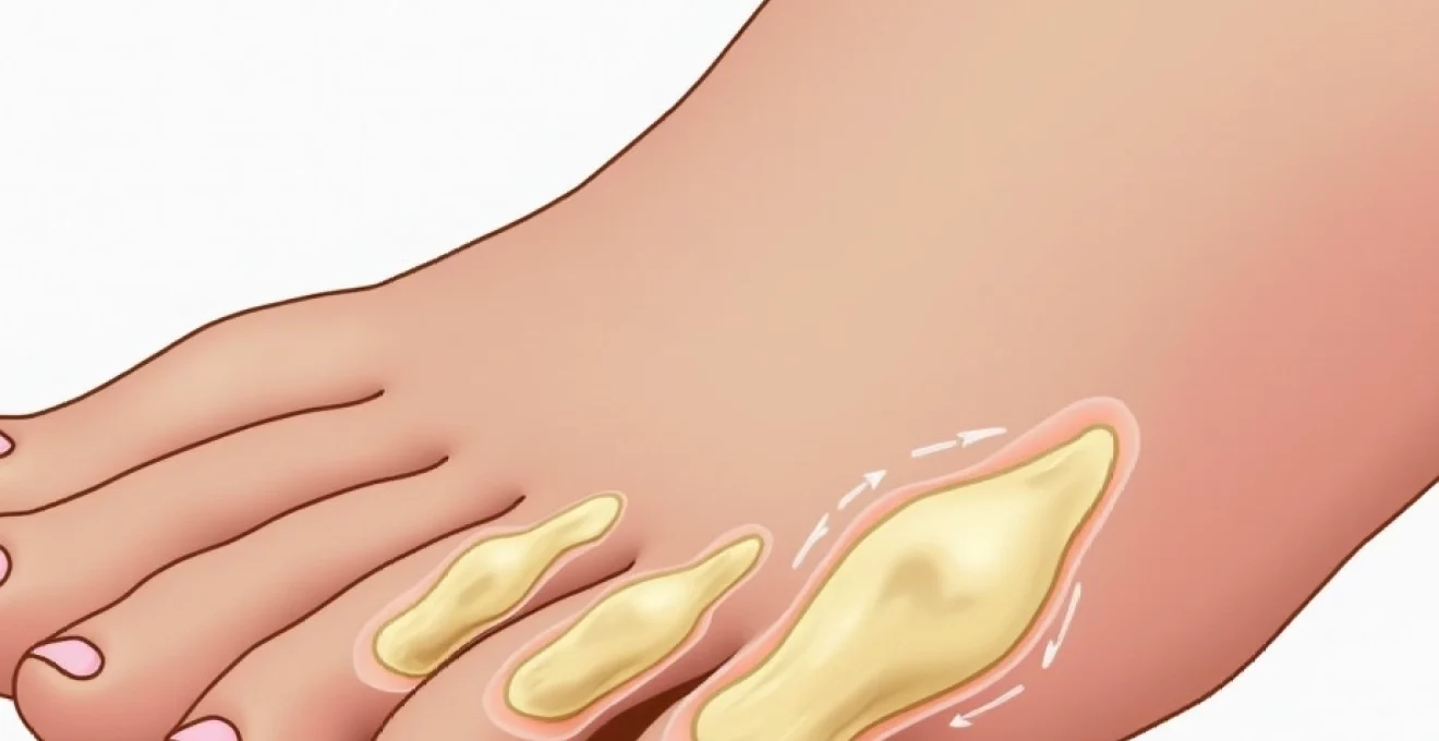
The development of a protruding big toe bone involves complex three-dimensional movements of multiple bone segments. The first metatarsal bone gradually shifts toward the midline of your foot, a movement called metatarsus primus adductus . Simultaneously, the proximal phalanx of your big toe deviates laterally toward the lesser toes, creating the classic bunion appearance that patients commonly observe.
This displacement pattern creates significant biomechanical alterations in your foot’s weight distribution. The normal tripod support system, consisting of the heel and both the first and fifth metatarsal heads, becomes compromised as the first ray loses its optimal positioning. Consequently, increased loading occurs across the lateral forefoot, potentially leading to secondary problems such as metatarsalgia and lesser toe deformities.
Bunion formation and medial eminence development
The visible bony prominence characteristic of bunions results from both bone displacement and reactive bone formation. As the first metatarsal shifts into its adducted position, the medial aspect of the metatarsal head becomes more prominent. Additionally, chronic pressure and friction from footwear stimulate osteoblastic activity, leading to the formation of exostotic bone growth along the medial eminence.
This reactive bone formation serves as your body’s attempt to strengthen the area experiencing increased mechanical stress. However, the resulting prominence often creates a cycle of worsening symptoms as the enlarged medial eminence encounters greater pressure from shoe wear, leading to inflammation of the overlying bursa and soft tissues. The combination of structural bone displacement and reactive bone growth creates the characteristic bunion deformity that becomes increasingly symptomatic over time.
Progressive joint subluxation in advanced cases
In severe hallux valgus deformities, the metatarsophalangeal joint may progress from simple angular deviation to frank subluxation or even dislocation. The sesamoid bones, small structures that normally glide beneath the first metatarsal head, become laterally displaced as the deformity worsens. This sesamoid displacement represents a critical milestone in bunion progression, as it indicates significant structural compromise that typically requires surgical intervention.
Joint subluxation creates additional biomechanical problems including hallux rigidus or stiffness of the big toe joint. The incongruent joint surfaces experience increased wear patterns, potentially leading to secondary osteoarthritis. Patients with subluxated joints often report difficulty with push-off during walking and may develop compensatory gait patterns that affect the entire kinetic chain from the foot to the lower back.
Biomechanical factors contributing to first ray protrusion
Pronation dysfunction and excessive foot rolling
Abnormal pronation patterns during the gait cycle represent a significant contributing factor to big toe bone protrusion. When your foot excessively pronates or remains pronated too long during the stance phase of walking, the first ray becomes hypermobile and unstable. This prolonged pronation causes the first metatarsal to plantarflex and invert excessively, creating an unstable foundation for the hallux.
The combination of first ray hypermobility and ground reaction forces during weight-bearing activities creates a perfect storm for bunion development. As your foot attempts to maintain stability during push-off, compensatory muscle activation patterns develop that may inadvertently contribute to hallux valgus progression. The peroneus longus muscle, which normally helps stabilize the first ray, may become overactive in response to the instability, potentially pulling the first metatarsal into further adduction.
Forefoot loading patterns during gait cycle
The distribution of forces across your forefoot during walking significantly influences the development of big toe bone protrusion. Normal gait patterns require approximately 40-60% of forefoot loading to occur through the first ray, with the remaining load distributed across the lesser metatarsals. When this loading pattern becomes altered due to structural or functional abnormalities, excessive stress may concentrate on specific areas.
Patients with cavus foot types or high-arched feet often demonstrate altered loading patterns that predispose to bunion formation. The rigid nature of cavus feet limits the foot’s ability to adapt to ground surfaces, concentrating forces on the first and fifth rays. This concentrated loading, combined with the typically tight plantar fascia associated with high arches, creates increased stress on the first metatarsophalangeal joint that may contribute to bunion development over time.
Achilles tendon tightness and compensatory mechanisms
Restricted ankle dorsiflexion due to Achilles tendon tightness creates cascading biomechanical effects that can contribute to big toe bone protrusion. When your ankle cannot achieve adequate dorsiflexion during the stance phase of gait, your foot must find alternative methods to clear the ground during swing phase. This compensation often occurs through increased pronation and midfoot mobility, potentially destabilizing the first ray.
The relationship between posterior muscle tightness and forefoot deformities demonstrates the interconnected nature of lower extremity biomechanics. Patients with equinus contractures frequently develop compensatory foot positioning that places increased stress on the medial column structures. This chronic stress pattern may accelerate the development of hallux valgus deformities, particularly when combined with other predisposing factors such as genetic susceptibility or inappropriate footwear choices.
Plantar fascia tension and first metatarsal mobility
The plantar fascia plays a crucial role in maintaining the stability and alignment of your foot’s medial column. This strong fibrous band connects your heel to the toes and helps support the longitudinal arch during weight-bearing activities. When plantar fascia tension becomes altered due to injury, tightness, or structural abnormalities, the resulting changes in foot mechanics can contribute to first ray instability.
Windlass mechanism dysfunction represents a specific biomechanical problem where the normal tightening of the plantar fascia during toe extension fails to adequately stabilize the medial column. This dysfunction allows excessive motion at the first ray, potentially contributing to the development of hallux valgus over time. Additionally, compensatory changes in muscle activation patterns around the first ray may develop as your foot attempts to maintain stability despite the compromised plantar fascia function.
Hereditary predisposition and genetic foot structure variations
Genetic factors play a substantial role in determining your susceptibility to developing protruding big toe bones. Research indicates that approximately 70% of individuals with bunions have a family history of the condition, suggesting strong hereditary influences on foot structure and biomechanics. The inheritance pattern appears complex, likely involving multiple genes that affect bone morphology, joint structure, ligament laxity, and muscle function.
Specific inherited foot characteristics that predispose to bunion formation include metatarsus adductus, where the forefoot naturally angles toward the midline of the body. Additionally, variations in metatarsal length patterns, particularly when the first metatarsal is shorter than the second, can create abnormal pressure distributions that contribute to hallux valgus development. Joint hypermobility syndrome, often inherited, increases the likelihood of developing bunions due to excessive ligament laxity throughout the foot and ankle complex.
The concept of foot type inheritance encompasses not only bone structure but also soft tissue characteristics such as tendon insertion points, muscle fiber composition, and ligament strength. These inherited variations create unique biomechanical signatures that may predispose certain individuals to specific foot pathologies. Understanding your family history of foot problems can provide valuable insights into your personal risk factors and help guide preventive strategies.
Studies show that individuals with a strong family history of bunions have a five times greater likelihood of developing the condition compared to those without genetic predisposition, highlighting the importance of early intervention and preventive measures.
Footwear-induced deformities and environmental pressure points
Narrow toe box compression and lateral force application
The relationship between footwear and big toe bone protrusion represents one of the most modifiable risk factors in bunion development. Shoes with narrow toe boxes create sustained lateral pressure on your big toe, gradually forcing it toward the lesser toes over months and years of wear. This chronic pressure particularly affects the soft tissue structures around the first metatarsophalangeal joint, including the joint capsule, ligaments, and surrounding musculature.
Fashion footwear often features pointed toe designs that concentrate forces on the hallux, creating a mechanical environment conducive to bunion formation. The sustained compression not only affects bone alignment but also compromises local circulation and tissue nutrition. Over time, these changes contribute to joint stiffness, capsular contracture, and the progressive angular deformity characteristic of hallux valgus. Women experience bunions at a rate approximately ten times higher than men, largely attributed to the frequent wearing of constrictive fashion footwear.
High-heel biomechanics and forefoot weight distribution
Elevated heel heights fundamentally alter the biomechanics of your foot and lower extremity during weight-bearing activities. When wearing high-heeled shoes, your body weight shifts anteriorly, increasing the load on your forefoot by up to 75% compared to flat shoes. This dramatic increase in forefoot loading places excessive stress on the first metatarsophalangeal joint, particularly when combined with the narrow toe box design typical of high-heeled footwear.
The biomechanical effects of high heels extend beyond simple weight redistribution. The elevated heel position shortens your calf muscles and Achilles tendon, creating compensatory changes throughout the kinetic chain. These adaptations may persist even when not wearing heels, potentially contributing to the development of functional equinus and its associated compensatory pronation patterns. Research indicates that regular high-heel wear correlates with increased bunion severity and accelerated progression of deformity.
Chronic shoe pressure and soft tissue adaptation
The human foot demonstrates remarkable adaptability to chronic environmental pressures, including those imposed by poorly fitting footwear. When shoes consistently apply pressure to specific areas of your foot, the underlying soft tissues undergo remodeling processes that may initially provide protection but ultimately contribute to deformity progression. The formation of calluses, corns, and bursal thickening represents your body’s attempt to adapt to chronic pressure points.
However, these adaptive changes often create a cycle of worsening symptoms and progressive deformity. Thickened skin and inflamed bursae occupy additional space within the shoe, creating even greater pressure and friction. The inflammatory response to chronic pressure can also affect nearby joint structures, contributing to capsular thickening, synovial inflammation, and eventual joint stiffness. Understanding these adaptation mechanisms helps explain why early intervention and proper footwear selection are crucial for preventing bunion progression.
Associated pathological conditions and secondary bone prominence
Several medical conditions can contribute to the development of protruding big toe bones, often accelerating the progression of bunion deformities beyond what might be expected from mechanical factors alone. Rheumatoid arthritis represents one of the most significant pathological contributors, as the chronic inflammatory process affects joint structures throughout the body, including the feet. The synovial inflammation characteristic of rheumatoid arthritis can destroy joint cartilage, weaken supporting ligaments, and create joint instability that predisposes to bunion formation.
Neuromuscular conditions such as cerebral palsy, stroke, or peripheral neuropathy can also contribute to big toe bone prominence through altered muscle balance and abnormal foot positioning. When the intrinsic muscles of your foot become weakened or paralyzed, the normal balance between flexor and extensor forces becomes disrupted. This imbalance can lead to progressive deformity development, including hallux valgus, particularly when combined with spasticity or abnormal muscle tone patterns.
Connective tissue disorders such as Ehlers-Danlos syndrome or Marfan syndrome create generalized joint hypermobility that significantly increases bunion risk. The excessive ligament laxity associated with these conditions allows abnormal joint motion that can accelerate deformity progression. Additionally, certain metabolic conditions affecting bone metabolism, such as hyperparathyroidism or osteomalacia, may contribute to bone deformity development through altered bone mineralization patterns.
Patients with inflammatory arthritis demonstrate bunion formation rates nearly three times higher than the general population, with more rapid progression and increased likelihood of requiring surgical intervention.
Post-traumatic bunions represent another important category where previous injuries to the foot or ankle create biomechanical alterations that predispose to later deformity development. Fractures involving the first ray, Lisfranc injuries, or ankle fractures that alter normal foot alignment can create abnormal stress patterns that contribute to bunion formation years after the initial injury. The concept of biomechanical compensation explains how the body adapts to post-traumatic changes, sometimes creating new problems in adjacent structures.
Clinical assessment techniques for protruding toe bone evaluation
Comprehensive evaluation of a protruding big toe bone requires systematic assessment of both structural and functional components. The clinical examination begins with careful observation of your foot in both weight-bearing and non-weight-bearing positions. This positional assessment helps distinguish between flexible and rigid deformities, which have different implications for treatment planning. Weight-bearing examination reveals the true extent of deformity under functional loading conditions, while non-weight-bearing assessment helps identify the reducibility of the deformity.
Range of motion testing at the first metatarsophalangeal joint provides crucial information about joint function and the presence of secondary arthritis. Normal dorsiflexion should measure approximately 65-70 degrees, with plantarflexion around 15-20 degrees. Significant limitations in either plane may indicate joint deterioration or soft tissue contracture that affects treatment options. Additionally, assessment of first ray mobility helps identify hypermobile or rigid first ray conditions that influence the biomechanical environment.
Radiographic evaluation remains the gold standard for assessing bunion severity and planning appropriate treatment. Weight-bearing X-rays provide accurate measurements of angular deformity, including the hallux valgus angle and intermetatarsal angle. These measurements help classify bunion severity and guide treatment recommendations. Advanced imaging such as CT scans or MRI may be necessary when evaluating complex deformities or suspected associated pathology such as osteonecrosis or occult fractures.
Functional assessment techniques include gait analysis and pressure distribution studies that reveal how the bunion affects your walking pattern and foot loading characteristics. Modern pressure measurement systems can identify areas of increased loading and help predict which patients may benefit from orthotic intervention. The integration of structural and functional assessment data provides a comprehensive understanding of how the protruding big toe bone affects overall foot function and quality of life, ultimately guiding the most appropriate treatment
approach that considers both the immediate symptoms and long-term foot health outcomes.
Biomechanical assessment tools such as pedobarography provide detailed information about pressure distribution patterns during walking and standing. These sophisticated measurement systems can identify abnormal loading patterns that may contribute to bunion progression or associated foot problems. The data obtained from pressure analysis helps clinicians understand how structural changes affect functional performance and can guide decisions about orthotic interventions or activity modifications.
Patient-reported outcome measures play an increasingly important role in clinical assessment, as they capture the subjective impact of the protruding big toe bone on daily activities and quality of life. Validated questionnaires such as the Manchester-Oxford Foot Questionnaire or the Foot and Ankle Ability Measure provide standardized methods for documenting symptom severity and functional limitations. These assessments help track treatment progress and ensure that interventions address the patient’s primary concerns and functional goals.
The integration of objective clinical findings with subjective patient experiences creates a comprehensive picture that guides treatment decision-making. Advanced assessment techniques, including three-dimensional foot scanning and dynamic radiography, continue to evolve and provide increasingly detailed information about foot structure and function. However, the fundamental principles of thorough history-taking, careful physical examination, and appropriate imaging remain the cornerstone of effective clinical evaluation for protruding big toe bone conditions.