Parkinson’s disease stiffness creates a distinctive sensation that patients frequently describe as feeling like their muscles have turned to concrete or lead. This rigidity represents one of the cardinal motor symptoms that define the condition, fundamentally altering how individuals experience movement and physical comfort. Unlike the temporary stiffness experienced after intense exercise or prolonged inactivity, Parkinsonian rigidity involves persistent muscle tension that can affect virtually any part of the body, from facial muscles to limbs and trunk.
The experience of rigidity in Parkinson’s disease extends far beyond simple muscle tightness. Patients often report feeling as though their joints have become rusted hinges, requiring tremendous effort to initiate even the simplest movements. This sensation can fluctuate throughout the day, often correlating with medication timing and various environmental factors. Understanding the multifaceted nature of Parkinsonian stiffness proves essential for both patients and healthcare providers in developing effective management strategies.
Bradykinesia and muscle rigidity: core manifestations of parkinsonian motor symptoms
The interplay between bradykinesia and muscle rigidity creates a complex symptom profile that distinguishes Parkinson’s disease from other movement disorders. Bradykinesia , characterised by slowness of movement, often compounds the effects of rigidity, making patients feel as though they’re moving through thick syrup or fighting against invisible resistance. This combination affects approximately 85% of Parkinson’s patients within the first five years of diagnosis, according to recent clinical studies.
Muscle rigidity in Parkinson’s disease occurs due to increased muscle tone throughout the entire range of motion, unlike spasticity where resistance varies with the speed of movement. Patients frequently describe this sensation as having muscles that refuse to relax, creating a constant state of tension that can be both uncomfortable and exhausting. The rigidity typically affects both agonist and antagonist muscle groups simultaneously, leading to what clinicians term “co-contraction.”
Lead-pipe rigidity versus cogwheel rigidity in clinical assessment
Lead-pipe rigidity produces a smooth, constant resistance throughout the entire range of motion, resembling the feel of bending a lead pipe. Patients experiencing this type of rigidity often describe their limbs as feeling heavy and unresponsive, requiring significant mental effort to coordinate even simple movements. This form of rigidity tends to be more prominent in advanced stages of the disease and can significantly impact functional abilities.
Cogwheel rigidity, conversely, creates a ratchet-like sensation with intermittent catches during passive movement. This phenomenon occurs when underlying tremor combines with lead-pipe rigidity, producing the characteristic jerky resistance pattern. Patients may notice this particularly during activities requiring smooth, continuous movements, such as stirring ingredients while cooking or brushing their hair.
Axial rigidity impact on postural control and truncal flexibility
Axial rigidity affects the muscles of the neck, trunk, and spine, profoundly impacting postural control and overall mobility. Patients often develop a characteristic stooped posture as truncal muscles become increasingly rigid, making it difficult to maintain an upright stance. This rigidity can contribute to the development of camptocormia, a severe forward flexion of the trunk that affects approximately 5-10% of Parkinson’s patients.
The loss of truncal flexibility also impacts respiratory function, as rigid intercostal muscles and diaphragmatic involvement can reduce lung capacity and breathing efficiency. Many patients report feeling short of breath during activities that previously posed no challenge, partly due to the increased effort required to expand the chest wall against rigid muscles.
Appendicular rigidity effects on limb movement coordination
Appendicular rigidity specifically affects the arms and legs, creating profound challenges in coordinating complex movements. Patients frequently describe their limbs as feeling disconnected from their intentions, requiring conscious effort to initiate movements that were once automatic. This type of rigidity particularly impacts fine motor control, making tasks such as buttoning clothes or handling utensils increasingly difficult.
The asymmetric nature of appendicular rigidity in early Parkinson’s disease often leads to compensatory movement patterns. Patients may unconsciously favour their less affected side, leading to muscle imbalances and potential secondary complications such as frozen shoulder or hip contractures over time.
Bradykinetic movement patterns in fine motor tasks
Fine motor tasks reveal the intricate relationship between bradykinesia and rigidity most clearly. Micrographia , the progressive decrease in handwriting size, exemplifies how these symptoms interact to impair precise movements. Patients often report that their handwriting starts normally but becomes increasingly small and cramped as they continue writing, reflecting the combined effects of rigidity and bradykinetic fatigue.
Dexterity tasks such as coin handling, card shuffling, or playing musical instruments become progressively more challenging as both symptoms advance. The combination creates a frustrating cycle where increased effort leads to greater fatigue, which in turn exacerbates both rigidity and bradykinesia.
Phenomenology of morning stiffness and Off-Period rigidity
Morning stiffness in Parkinson’s disease represents one of the most challenging aspects of daily symptom management. Unlike morning stiffness associated with arthritis or aging, Parkinsonian morning rigidity can be severe enough to prevent patients from getting out of bed without assistance. This phenomenon occurs due to the natural decline in dopamine levels during sleep, combined with the wearing-off effects of medications taken the previous evening.
Patients often describe waking up feeling as though they’ve been “frozen overnight,” with muscles that seem locked in position. Simple tasks such as turning over in bed, sitting up, or placing feet on the floor can require significant effort and time. This morning rigidity typically improves within 30-90 minutes after taking the first dose of levodopa, highlighting the medication-dependent nature of symptom control in Parkinson’s disease.
Levodopa Wearing-Off effects on muscle tone regulation
The wearing-off phenomenon creates predictable patterns of rigidity that many patients learn to anticipate and manage. As levodopa levels decline in the bloodstream, muscle tone gradually increases, often beginning 3-4 hours after the last dose. Patients frequently describe this as feeling like their body is “tightening up” or “seizing up,” with movements becoming progressively more difficult and effortful.
Advanced patients may experience what clinicians term “dose failures,” where rigidity returns abruptly rather than gradually. These episodes can be particularly distressing, as patients may find themselves suddenly unable to move normally, sometimes in the middle of activities or social situations.
Nocturnal akinesia and early morning mobility challenges
Nocturnal akinesia encompasses the severe rigidity and movement difficulties that occur during nighttime hours. Patients often report being unable to turn over in bed, adjust their position, or get up to use the bathroom without significant struggle. This can lead to sleep fragmentation and increased daytime fatigue, creating a cycle that exacerbates other Parkinson’s symptoms.
Early morning mobility challenges extend beyond simple stiffness to include complex difficulties with bed mobility, transfers, and initial ambulation. Many patients develop specific strategies for managing these challenges, such as keeping medication bedside for early morning administration or using assistive devices to help with bed mobility.
Dystonic rigidity during medication fluctuations
Dystonic rigidity represents a particularly uncomfortable manifestation that can occur during off-periods or as part of wearing-off phenomena. This involves sustained muscle contractions that create twisted, abnormal postures, often affecting the feet, hands, or neck. Patients describe these episodes as intensely uncomfortable, with muscles that feel locked in unnatural positions.
Morning dystonia, particularly affecting the feet and toes, affects approximately 50% of Parkinson’s patients and can be one of the most painful aspects of the condition. The sensation is often described as cramping combined with rigidity, making weight-bearing and walking extremely uncomfortable until medications take effect.
Temperature sensitivity and environmental rigidity triggers
Many Parkinson’s patients report increased rigidity in response to environmental factors, particularly cold temperatures. This phenomenon, sometimes called “cold-induced rigidity,” can make winter months particularly challenging. Patients often notice that their symptoms worsen significantly when exposed to cold air, requiring additional layers of clothing or heated environments to maintain optimal mobility.
Stress and emotional tension can also trigger increased rigidity episodes. The body’s natural stress response appears to exacerbate muscle tension in Parkinson’s patients, creating a feedback loop where anxiety about movement difficulties leads to increased rigidity, which in turn increases anxiety levels.
Anatomical distribution patterns of parkinsonian stiffness
The distribution of rigidity in Parkinson’s disease follows characteristic patterns that reflect the underlying pathophysiology of the condition. Initial rigidity typically begins unilaterally, most commonly affecting the shoulder and arm on one side before progressing to involve other muscle groups. This asymmetric onset remains a hallmark of idiopathic Parkinson’s disease, helping differentiate it from other parkinsonian syndromes that may present with more symmetric involvement.
As the disease progresses, rigidity spreads in a predictable pattern, generally moving from the initially affected side to involve bilateral upper extremities, then progressing to affect axial muscles and lower extremities. Research indicates that approximately 70% of patients will develop bilateral rigidity within three years of initial symptom onset, though the originally affected side typically remains more severely impacted throughout the disease course.
The severity and distribution of rigidity can vary significantly based on the predominant disease subtype. Tremor-dominant patients often experience less severe rigidity compared to those with the akinetic-rigid subtype, where rigidity may be the most prominent and disabling feature. Understanding these patterns helps both patients and healthcare providers anticipate symptom progression and plan appropriate interventions.
The progression of rigidity in Parkinson’s disease follows anatomical patterns that can help predict functional challenges and guide therapeutic interventions, making early recognition and documentation crucial for optimal management.
Subjective patient experiences of rigidity and movement restriction
Patient descriptions of Parkinsonian rigidity reveal the profound impact this symptom has on daily life and overall quality of life. Many individuals struggle to articulate the unique sensation of rigidity, often resorting to vivid metaphors and analogies to convey their experience. Common descriptions include feeling like a “rusty robot,” having muscles made of “concrete,” or moving through “invisible quicksand.” These descriptions highlight the alien nature of the sensation for patients who previously enjoyed fluid, effortless movement.
The psychological impact of rigidity extends beyond physical discomfort to affect self-perception and confidence. Many patients report feeling betrayed by their bodies, describing a disconnect between their mental intentions and physical capabilities. This phenomenon can lead to increased anxiety around movement, creating what some researchers term “kinesiophobia” – fear of movement that can actually worsen rigidity through increased muscle tension.
Frozen shoulder syndrome in parkinson’s disease patients
Frozen shoulder, or adhesive capsulitis, occurs at significantly higher rates in Parkinson’s patients, affecting approximately 25-40% of individuals compared to 2-5% in the general population. The combination of reduced arm swing, prolonged immobility, and intrinsic rigidity creates ideal conditions for shoulder capsule contracture development. Patients often describe a gradual onset of shoulder pain and stiffness that differs from general Parkinsonian rigidity.
The progression of frozen shoulder in Parkinson’s disease can be particularly challenging because it layers additional movement restriction on top of existing rigidity. Patients frequently report being unable to distinguish between symptoms caused by their Parkinson’s disease and those related to the shoulder condition, making treatment more complex and potentially delaying appropriate interventions.
Facial masking and orofacial rigidity sensations
Facial masking , or hypomimia, results from rigidity affecting the muscles of facial expression, creating a mask-like appearance that can significantly impact social interactions. Patients often report feeling like their face has become “frozen” or “wooden,” making it difficult to convey emotions naturally. This can be particularly frustrating during social situations where facial expressions play a crucial role in communication.
Orofacial rigidity extends beyond facial expression to affect chewing, swallowing, and speech production. Many patients notice that their jaw feels tight or locked, making it difficult to open their mouth fully for eating or dental care. The muscles involved in speech production can also become rigid, contributing to the characteristic soft, monotone speech pattern associated with Parkinson’s disease.
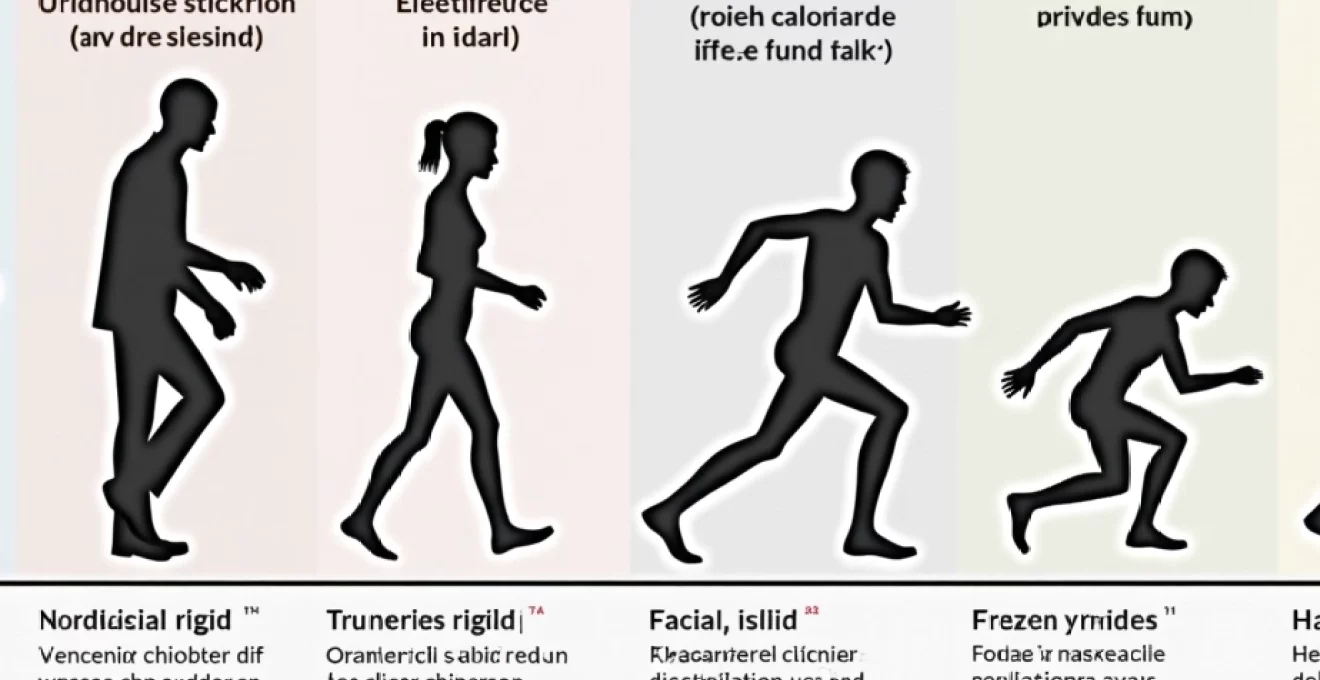
Gait freezing episodes and lower extremity stiffness
Gait freezing represents one of the most distinctive and potentially dangerous manifestations of lower extremity rigidity in Parkinson’s disease. Patients describe the sensation as having their feet “glued to the ground” or feeling like they’re trying to walk through deep mud. These episodes typically occur when initiating walking, turning, or approaching narrow spaces such as doorways.
The relationship between lower extremity rigidity and freezing episodes appears complex, with rigidity potentially contributing to the mechanical aspects of freezing while other factors influence the neurological components. Patients often develop compensatory strategies such as visual cues, rhythmic counting, or specific movement patterns to overcome freezing episodes and associated rigidity.
Handwriting changes and Micrographia-Related tension
Handwriting changes in Parkinson’s disease reflect the complex interaction between rigidity, bradykinesia, and tremor. Patients consistently report increased muscle tension while writing, describing the need to grip pens more tightly and apply greater pressure to paper. This increased effort often leads to hand cramping and fatigue during writing tasks that were previously effortless.
The progressive nature of micrographia creates additional frustration as patients notice their handwriting becoming increasingly difficult to read. Many individuals report consciously trying to write larger letters, only to find their writing becoming progressively smaller despite their efforts, highlighting the involuntary nature of these motor changes.
Dopaminergic medication response patterns in rigidity management
The response of rigidity to dopaminergic medications provides crucial insights into both the underlying pathophysiology of Parkinson’s disease and optimal treatment strategies. Levodopa remains the gold standard for rigidity management, typically providing significant improvement within 30-60 minutes of administration in most patients. However, the response pattern can vary considerably between individuals and may change over the course of the disease progression.
Initial medication response often produces dramatic improvements in rigidity, with patients describing feeling like they’ve been “unlocked” or “freed” from their physical constraints. This honeymoon period, typically lasting 2-5 years, allows many patients to maintain near-normal function with relatively simple medication regimens. However, as the disease progresses, medication response patterns become more complex, requiring careful timing and dosing adjustments to maintain optimal rigidity control.
The development of motor fluctuations introduces additional complexity to rigidity management. Patients begin to experience predictable patterns of symptom return as medication effects wear off, often describing a gradual increase in muscle tension and stiffness that signals the need for their next dose. Understanding these patterns allows patients to time activities around medication schedules and helps healthcare providers optimize dosing regimens.
Effective rigidity management requires understanding individual response patterns to dopaminergic medications, as these patterns guide timing strategies and dosing optimizations throughout disease progression.
Progressive stiffness stages throughout hoehn and yahr scale advancement
The evolution of rigidity throughout different stages of Parkinson’s disease follows predictable patterns that correlate with the widely-used Hoehn and Yahr staging system. In Stage 1, rigidity typically affects one side of the body and may be subtle enough that patients attribute symptoms to aging, overuse, or other conditions. During this early stage, rigidity often manifests as shoulder stiffness, reduced arm swing during walking, or difficulty with fine motor tasks on the affected side.
Stage 2 progression introduces bilateral involvement, though symptoms remain manageable without significant impact on daily activities. Patients at this stage often notice increased effort required for movements that were previously automatic, such as getting up from chairs, turning in bed, or performing sequential movements. The rigidity becomes more noticeable to family members and friends, often prompting medical evaluation for the first time.
Advanced stages (3-5) of Parkinson’s disease present increasingly severe rigidity that significantly impacts functional independence. Stage 3 patients typically experience rigidity that interferes with balance and increases fall risk, while maintaining general independence. By Stages 4 and 5, rigidity can become so severe that it contributes to complete dependency for activities of daily living, requiring comprehensive care approaches that address both motor symptoms and their secondary complications.
The progression of rigidity through these stages isn’t necessarily linear or uniform across all muscle groups. Some patients
may experience more rapid progression in axial muscles while limb rigidity remains relatively stable, or vice versa. This variability underscores the importance of individualized assessment and treatment planning throughout the disease course.
Understanding the relationship between Hoehn and Yahr progression and rigidity patterns helps patients and families prepare for future challenges while maintaining realistic expectations about disease trajectory. Early intervention with physical therapy, exercise programs, and optimal medication management can potentially slow rigidity progression and preserve functional capacity longer than would occur with disease progression alone.
The impact of rigidity on quality of life measures also correlates strongly with disease stage advancement. While early-stage patients may report minimal functional impact from rigidity, advanced-stage individuals often identify muscle stiffness as one of their most disabling symptoms. This progression highlights the critical importance of proactive rigidity management strategies that evolve with disease severity to maintain optimal function and comfort throughout the Parkinson’s journey.