Cervical disc osteophyte complexes at the C5-6 and C6-7 levels represent some of the most challenging degenerative pathologies encountered in spinal medicine. These conditions, characterised by the simultaneous presence of disc herniation and bony spur formation, create a complex interplay of mechanical and neurological dysfunction that can significantly impact patient quality of life. The C5-6 and C6-7 segments bear the brunt of cervical spine loading during daily activities, making them particularly susceptible to degenerative changes that progress over time.
Modern understanding of disc osteophyte complexes has evolved considerably, with advanced imaging techniques revealing the intricate relationship between disc degeneration, osteophyte formation, and neural compression. These pathological processes do not occur in isolation but rather represent a cascade of biomechanical adaptations that the cervical spine undergoes in response to chronic stress and age-related changes. Recognition of the multifactorial nature of these conditions is essential for developing effective treatment strategies that address both symptomatic relief and long-term spinal health.
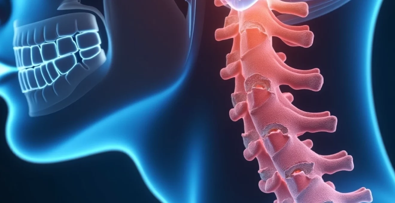
Cervical spine anatomy and degenerative pathophysiology at C5-6 and C6-7 levels
The cervical spine’s unique anatomical configuration makes it particularly vulnerable to degenerative changes, especially at the C5-6 and C6-7 motion segments. These levels experience the greatest range of motion during flexion and extension activities, subjecting the intervertebral discs and adjacent vertebral bodies to repetitive mechanical stress. The transition from the mobile cervical spine to the relatively fixed thoracic spine at C7-T1 creates additional biomechanical challenges that contribute to accelerated degeneration at these critical junctions.
Understanding the progression from normal anatomy to pathological changes requires appreciation of the complex interplay between multiple structures. The uncovertebral joints, unique to the cervical spine, play a crucial role in guiding motion whilst simultaneously being prone to arthritic changes that contribute to foraminal stenosis. As these joints degenerate, they lose their ability to maintain proper segmental alignment, leading to abnormal stress distribution across the disc space and facet joints.
Intervertebral disc structure and annulus fibrosus degeneration mechanisms
The intervertebral disc consists of three distinct components: the nucleus pulposus, annulus fibrosus, and cartilaginous endplates. In healthy discs, the nucleus pulposus maintains high water content and proteoglycan concentration, providing hydraulic support that distributes loads evenly across the annulus fibrosus. The annulus fibrosus comprises concentric lamellae of collagen fibres arranged in alternating orientations, creating a sophisticated containment system for the nucleus whilst allowing controlled motion between vertebrae.
Degenerative changes typically begin with biochemical alterations in the nucleus pulposus, including decreased proteoglycan synthesis and increased matrix metalloproteinase activity. These changes lead to progressive dehydration and loss of disc height, fundamentally altering the biomechanics of the motion segment. As the nucleus loses its ability to maintain intradiscal pressure, abnormal stresses are transferred to the annulus fibrosus, initiating a cascade of structural failures that eventually result in disc bulging, herniation, and associated osteophyte formation.
Osteophyte formation through wolff’s law and mechanical stress distribution
Osteophyte development represents the body’s attempt to stabilise degenerating motion segments through increased surface area for load distribution. According to Wolff’s Law, bone remodels in response to mechanical stress, and osteophytes form as a direct consequence of altered loading patterns caused by disc degeneration. The process begins with microtears in the annulus fibrosus and adjacent vertebral endplates, triggering an inflammatory response that stimulates osteoblastic activity at the vertebral margins.
The morphology and location of osteophytes provide valuable insights into the underlying degenerative process. Anterior osteophytes typically develop in response to flexion-based loading patterns, whilst posterior osteophytes are more commonly associated with extension-based activities and tend to cause greater clinical significance due to their proximity to neural structures. The size and extent of osteophyte formation generally correlate with the severity and duration of the degenerative process, though individual variations in bone metabolism can influence this relationship.
Neural foraminal stenosis and spinal canal narrowing biomechanics
Neural foraminal stenosis represents one of the most clinically significant consequences of disc osteophyte complexes at C5-6 and C6-7 levels. The neural foramen, bounded by the uncovertebral joint anteriorly, the facet joint posteriorly, and the intervertebral disc inferiorly, becomes progressively narrowed as degenerative changes advance. This three-dimensional narrowing process affects both the static and dynamic dimensions of the foramen, with symptoms often exacerbated by cervical extension and ipsilateral rotation.
Spinal canal stenosis in the cervical region differs significantly from lumbar stenosis due to the smaller cross-sectional area of the cervical canal and the presence of the cervical enlargement. Even modest degrees of canal narrowing can result in significant myelopathic symptoms, particularly when combined with dynamic compression during neck motion. The concept of functional stenosis becomes particularly relevant in cervical pathology, where symptoms may occur despite apparently adequate static measurements on imaging studies.
Cervical lordosis alterations and segmental motion dysfunction
Loss of cervical lordosis represents both a consequence and a contributing factor to disc osteophyte complex formation. As disc height decreases and osteophytes develop, the normal cervical curvature becomes straightened or even reversed, leading to significant alterations in sagittal balance and load distribution. This kyphotic deformity increases the mechanical advantage of flexion moments, accelerating further degenerative changes and creating a self-perpetuating cycle of dysfunction.
Segmental motion analysis reveals complex patterns of hypermobility and hypomobility that develop as the spine attempts to compensate for dysfunctional segments. Adjacent level changes often occur as a result of altered kinematics, with increased motion demands placed on healthier segments to maintain overall cervical function. Understanding these compensatory mechanisms is crucial for predicting treatment outcomes and planning appropriate interventions that address the entire kinetic chain rather than isolated pathological segments.
Advanced imaging techniques for disc osteophyte complex assessment
Modern diagnostic imaging has revolutionised the assessment of cervical disc osteophyte complexes, providing unprecedented detail regarding both morphological changes and functional implications. The complexity of cervical anatomy requires a multimodal imaging approach that combines the strengths of different techniques to provide comprehensive evaluation of osseous, soft tissue, and neural elements. Advanced imaging protocols have evolved to address specific clinical questions whilst minimising radiation exposure and providing clinically relevant information for treatment planning.
The evolution from conventional radiography to sophisticated cross-sectional imaging has transformed our understanding of cervical pathology. Modern imaging techniques allow for precise measurement of canal dimensions, assessment of neural compromise, and evaluation of dynamic instability patterns that were previously undetectable. This enhanced diagnostic capability has significant implications for both surgical planning and prognostic assessment, enabling more targeted interventions and improved patient outcomes.
MRI T2-Weighted sagittal sequences and disc signal intensity analysis
T2-weighted sagittal sequences represent the cornerstone of cervical disc evaluation, providing exquisite detail regarding disc hydration status and morphological changes. The high water content of healthy discs creates characteristic hyperintense signal on T2-weighted images, whilst degenerative changes result in progressive signal loss that correlates with biochemical alterations within the nucleus pulposus. Standardised grading systems, such as the Pfirrmann classification, enable objective assessment of degenerative severity and facilitate longitudinal monitoring of disease progression.
Advanced MRI sequences, including T2 mapping and diffusion-weighted imaging, provide quantitative assessment of disc composition and metabolic activity. These techniques offer valuable insights into early degenerative changes that precede morphological alterations visible on conventional sequences. Quantitative analysis of disc signal intensity changes has shown promise as a biomarker for predicting treatment response and identifying patients at risk for accelerated degeneration.
CT myelography with multiplanar reconstruction for bony detail visualisation
CT myelography remains the gold standard for evaluating complex cervical pathology involving both osseous and soft tissue components. The combination of intrathecal contrast administration with high-resolution CT imaging provides exceptional detail regarding neural compression patterns and the relationship between disc material and osteophytic spurring. Multiplanar reconstruction capabilities allow for precise assessment of foraminal dimensions and identification of subtle compression patterns that may be missed on axial images alone.
Modern CT myelography protocols incorporate isotropic voxel acquisition and advanced reconstruction algorithms that enable detailed evaluation of neural foramen geometry and spinal cord morphology. The ability to differentiate between soft disc herniation and hard osteophytic compression has significant treatment implications, particularly when considering surgical approaches and expected outcomes. Dynamic CT myelography , performed in flexion and extension positions, provides additional information regarding positional changes in neural compression that may guide activity modifications and treatment planning.
Flexion-extension radiographs for dynamic instability evaluation
Dynamic radiographic evaluation remains essential for assessing segmental stability and identifying pathological motion patterns in patients with cervical disc osteophyte complexes. Flexion-extension radiographs provide quantitative assessment of translational and angular motion at each cervical segment, enabling identification of hypermobility patterns that may contribute to symptom generation. Standardised measurement techniques, including assessment of sagittal translation and angular motion, provide objective criteria for defining instability.
The interpretation of dynamic radiographs requires careful consideration of patient positioning, effort, and pain limitations that may influence motion measurements. Subtle instability patterns may only become apparent when patients are able to achieve full range of motion without protective muscle guarding. Advanced analysis techniques, including computer-assisted measurement systems, improve the accuracy and reproducibility of instability assessment whilst reducing inter-observer variability in interpretation.
Gradient echo MRI sequences for cerebrospinal fluid flow assessment
Phase-contrast and time-resolved gradient echo sequences enable non-invasive assessment of cerebrospinal fluid dynamics and identification of flow disturbances associated with cervical stenosis. These techniques provide functional information regarding the severity of spinal canal compromise and may help differentiate between significant and incidental imaging findings. CSF flow analysis has shown particular value in assessing patients with myelopathic symptoms where conventional imaging findings may appear disproportionate to clinical presentation.
Advanced MRI techniques, including diffusion tensor imaging and functional connectivity analysis, are emerging as research tools for understanding the neurological impact of cervical pathology. These methods provide insights into white matter integrity and neural network function that may help predict treatment outcomes and identify optimal intervention timing. Whilst currently primarily research-based, these techniques may eventually find clinical application in complex cases where conventional imaging and clinical assessment provide conflicting information.
Clinical manifestations and neurological examination findings
The clinical presentation of cervical disc osteophyte complexes at C5-6 and C6-7 levels encompasses a broad spectrum of symptoms ranging from localised neck pain to complex myeloradiculopathy syndromes. Understanding the anatomical basis for symptom generation requires detailed knowledge of nerve root distributions and the relationship between specific pathological changes and clinical manifestations. The C6 and C7 nerve roots, most commonly affected by pathology at these levels, innervate critical upper extremity functions, making accurate diagnosis essential for appropriate treatment planning.
Symptom onset may be insidious, with gradual progression over months to years, or acute following trauma or specific inciting events. Many patients describe episodic symptoms that fluctuate with activity level and neck positioning, reflecting the dynamic nature of neural compression in the cervical spine. The complexity of symptom patterns often requires careful differentiation from other conditions, including peripheral nerve entrapments, rotator cuff pathology, and thoracic outlet syndrome.
Neurological examination findings in patients with C5-6 and C6-7 disc osteophyte complexes typically reflect the specific nerve roots involved and the degree of compression present. C6 radiculopathy commonly presents with weakness in elbow flexion (biceps), wrist extension, and thumb extension, accompanied by sensory changes in the thumb and index finger. C7 radiculopathy characteristically affects elbow extension (triceps), wrist flexion, and finger extension, with sensory symptoms involving the middle finger and dorsal hand.
The distinction between radiculopathy and myelopathy becomes crucial when evaluating patients with cervical disc osteophyte complexes, as the treatment implications differ significantly. Myelopathic symptoms may include gait disturbances, fine motor dysfunction, and upper motor neuron signs such as hyperreflexia and pathological reflexes. Early recognition of myelopathy is essential, as delay in treatment may result in irreversible neurological deficit. Subtle myelopathic signs may include difficulty with fine motor tasks such as buttoning clothes or writing, balance problems, and subjective feelings of leg weakness or stiffness.
The key to successful management lies in accurate assessment of the relationship between imaging findings and clinical symptoms, as anatomical abnormalities may exist without causing significant functional impairment.
Provocative testing during clinical examination can help localise pathology and assess the functional significance of imaging findings. Spurling’s test, involving neck extension with rotation and lateral flexion toward the symptomatic side, may reproduce radicular symptoms by further narrowing the neural foramen. The shoulder abduction relief sign, where elevation of the affected arm above the head reduces symptoms, suggests nerve root compression as the primary pain generator. These clinical tests, whilst not pathognomonic, provide valuable information when interpreted in conjunction with imaging findings.
Pain patterns associated with cervical disc osteophyte complexes often extend beyond the classic dermatome distributions, reflecting the complex nature of referred pain and the involvement of multiple pain-generating structures. Facet joint arthropathy, muscle spasm, and myofascial dysfunction commonly accompany disc pathology, creating overlapping symptom patterns that can complicate diagnosis. Understanding these pain mechanisms is essential for developing comprehensive treatment plans that address all contributing factors rather than focusing solely on the primary structural pathology.
Conservative management strategies and Evidence-Based interventions
Conservative management remains the first-line treatment approach for most patients with cervical disc osteophyte complexes, with success rates ranging from 60-90% depending on symptom severity and duration. The multifaceted nature of these conditions requires a comprehensive treatment approach that addresses inflammation, mechanical dysfunction, and functional impairment simultaneously. Evidence-based protocols emphasise the importance of early intervention, patient education, and progressive functional restoration to optimise outcomes and prevent chronicity.
Pharmacological interventions form the foundation of acute symptom management, with non-steroidal anti-inflammatory drugs (NSAIDs) providing effective pain relief and anti-inflammatory effects. Neuropathic pain medications, including gabapentin and pregabalin, demonstrate particular efficacy for radicular symptoms associated with nerve root compression. Muscle relaxants may provide short-term benefit during acute exacerbations, though their long-term use should be limited due to sedating effects and dependency potential.
Physical therapy represents a cornerstone of conservative management, with exercise-based interventions demonstrating significant efficacy in reducing pain and improving functional outcomes. Cervical stabilisation exercises focus on strengthening the deep cervical flexors and improving neuromuscular control, whilst mobility exercises address restrictions in range of motion and tissue extensibility. Manual therapy techniques , including joint mobilisation and soft tissue techniques, provide additional benefits when combined with exercise interventions.
- Cervical collar use for acute symptom relief and activity modification
- Heat and cold therapy applications for inflammation control
- Ergonomic modifications to reduce occupational stress factors
- Patient education regarding proper body mechanics and posture
Interventional pain management techniques offer additional options for patients who do not respond adequately to initial conservative measures. Cervical epidural steroid injections provide targeted anti-inflammatory treatment directly to the affected nerve roots, with success rates of 60-80% for appropriately selected patients. Selective nerve root blocks serve both diagnostic and therapeutic purposes, helping to confirm the pain generator whilst providing temporary symptom relief that may facilitate participation in rehabilitative activities.
The timing and selection of interventional procedures requires careful consideration of patient factors, symptom severity, and functional goals. Cervical facet joint injections may be appropriate for patients with predominantly axial neck pain and evidence of facet arthropathy on imaging studies. Radiofrequency ablation procedures offer longer-lasting pain relief for patients who respond positively to diagnostic facet blocks, though careful patient selection is essential to optimise outcomes
Surgical treatment options and procedural considerations
Surgical intervention becomes necessary when conservative management fails to provide adequate symptom relief or when progressive neurological deficits develop. The decision to proceed with surgery requires careful consideration of multiple factors, including symptom severity, functional impairment, imaging findings, and patient expectations. Modern surgical techniques offer several options for addressing cervical disc osteophyte complexes, each with specific indications, advantages, and potential complications that must be thoroughly discussed during the informed consent process.
The primary goals of surgical treatment include neural decompression, restoration of spinal alignment, and prevention of further degenerative changes. Achieving these objectives often requires removal of both soft disc material and hard osteophytic bone, making surgical planning more complex than isolated disc herniations. The choice of surgical approach depends on the predominant pathology, number of levels involved, sagittal alignment, and surgeon experience. Anterior cervical approaches remain the gold standard for most cervical disc osteophyte complexes, offering excellent visualization of the disc space and neural elements whilst allowing for immediate reconstruction and stabilisation.
Anterior cervical discectomy and fusion (ACDF) represents the most commonly performed procedure for cervical disc osteophyte complexes at C5-6 and C6-7 levels. This technique involves complete removal of the intervertebral disc and associated osteophytes through an anterior approach, followed by insertion of a bone graft or interbody cage to maintain disc height and promote fusion. The procedure provides excellent decompression of both the spinal canal and neural foramina whilst restoring sagittal alignment and preventing recurrent pathology. Long-term fusion rates exceed 95% with modern techniques, though adjacent level degeneration remains a concern with rates of 10-15% requiring additional surgery within ten years.
Cervical disc arthroplasty has emerged as an alternative to fusion for appropriately selected patients with single-level pathology and preserved facet joint integrity. Motion-preserving devices aim to maintain segmental kinematics whilst providing neural decompression, theoretically reducing the risk of adjacent level degeneration. However, the presence of significant osteophytic spurring and facet arthropathy often limits the applicability of arthroplasty, as these conditions typically require fusion for optimal outcomes. Strict patient selection criteria must be applied to ensure appropriate candidacy for motion preservation procedures.
Posterior approaches, including laminectomy and laminoplasty, are reserved for patients with predominantly posterior compression or multilevel stenosis where anterior reconstruction would be technically challenging. These techniques provide indirect decompression by expanding the spinal canal diameter, though they do not directly address anterior pathology such as disc herniations or anterior osteophytes. The choice between laminectomy and laminoplasty depends on the extent of pathology and the need to preserve posterior cervical stability. Laminoplasty offers the advantage of maintaining some degree of posterior stability whilst providing adequate decompression for multilevel disease.
Minimally invasive techniques have gained popularity for selected cases, offering potential advantages including reduced tissue trauma, shorter operative times, and faster recovery. Percutaneous endoscopic discectomy and microscopic approaches through smaller incisions can provide adequate decompression for soft disc herniations, though their utility in managing complex osteophytic pathology remains limited. The learning curve for minimally invasive techniques is substantial, and appropriate case selection is crucial to avoid complications and suboptimal outcomes.
The key to successful surgical outcomes lies in matching the appropriate procedure to the specific pathological anatomy whilst considering patient factors that may influence healing and recovery.
Intraoperative monitoring techniques, including somatosensory and motor evoked potentials, provide real-time assessment of spinal cord function during decompression procedures. These monitoring modalities are particularly valuable when operating near the spinal cord or when significant canal stenosis is present. The ability to detect neurological changes during surgery allows for immediate corrective action and may reduce the risk of postoperative neurological complications. However, monitoring is not infallible, and careful surgical technique remains paramount in preventing injury to neural structures.
Postoperative management protocols have evolved to emphasise early mobilisation and functional restoration whilst protecting the surgical construct during healing. Modern fusion techniques often allow for immediate mobilisation without external orthoses, though patient-specific factors may necessitate temporary collar use. Physical therapy typically begins within the first few weeks following surgery, focusing initially on range of motion and progressing to strengthening exercises as fusion consolidates. Patient education regarding activity restrictions and warning signs of complications remains crucial for optimising outcomes and preventing adverse events.
Complications associated with cervical spine surgery include both general surgical risks and procedure-specific concerns. Adjacent level degeneration represents the most significant long-term complication of fusion procedures, though its clinical significance varies considerably between patients. Pseudarthrosis rates have decreased dramatically with improved surgical techniques and graft materials, though smoking and other patient factors continue to influence fusion success. Neural injury, though rare with experienced surgeons, represents the most feared complication and emphasises the importance of appropriate surgical training and case selection.
The economic implications of surgical treatment must be considered in the context of healthcare resource allocation and patient financial burden. Whilst surgical intervention involves significant upfront costs, successful procedures often result in substantial long-term savings through reduced healthcare utilisation, improved work productivity, and enhanced quality of life. Cost-effectiveness analyses consistently demonstrate favourable outcomes for surgical treatment of appropriate candidates, though careful patient selection remains essential to optimise resource utilisation. The development of value-based care models emphasises the importance of achieving meaningful clinical outcomes rather than simply performing procedures.