Double-J stent removal marks a significant milestone in urological recovery, yet many patients find themselves unprepared for the discomfort that follows this seemingly straightforward procedure. While the extraction itself typically takes only minutes, the subsequent pain and urinary symptoms can persist for several days, causing considerable anxiety amongst those unfamiliar with normal recovery patterns. Understanding what constitutes typical post-removal discomfort versus warning signs requiring immediate medical attention becomes crucial for patient wellbeing and peace of mind.
Research indicates that approximately 27% of patients experience increased pain in the hours following ureteral stent removal, with certain demographic groups showing higher susceptibility. Female patients demonstrate a 2.41 times greater likelihood of experiencing post-removal pain, whilst younger individuals also face elevated risk compared to their older counterparts. These statistics highlight the importance of personalised patient counselling and preparation strategies.
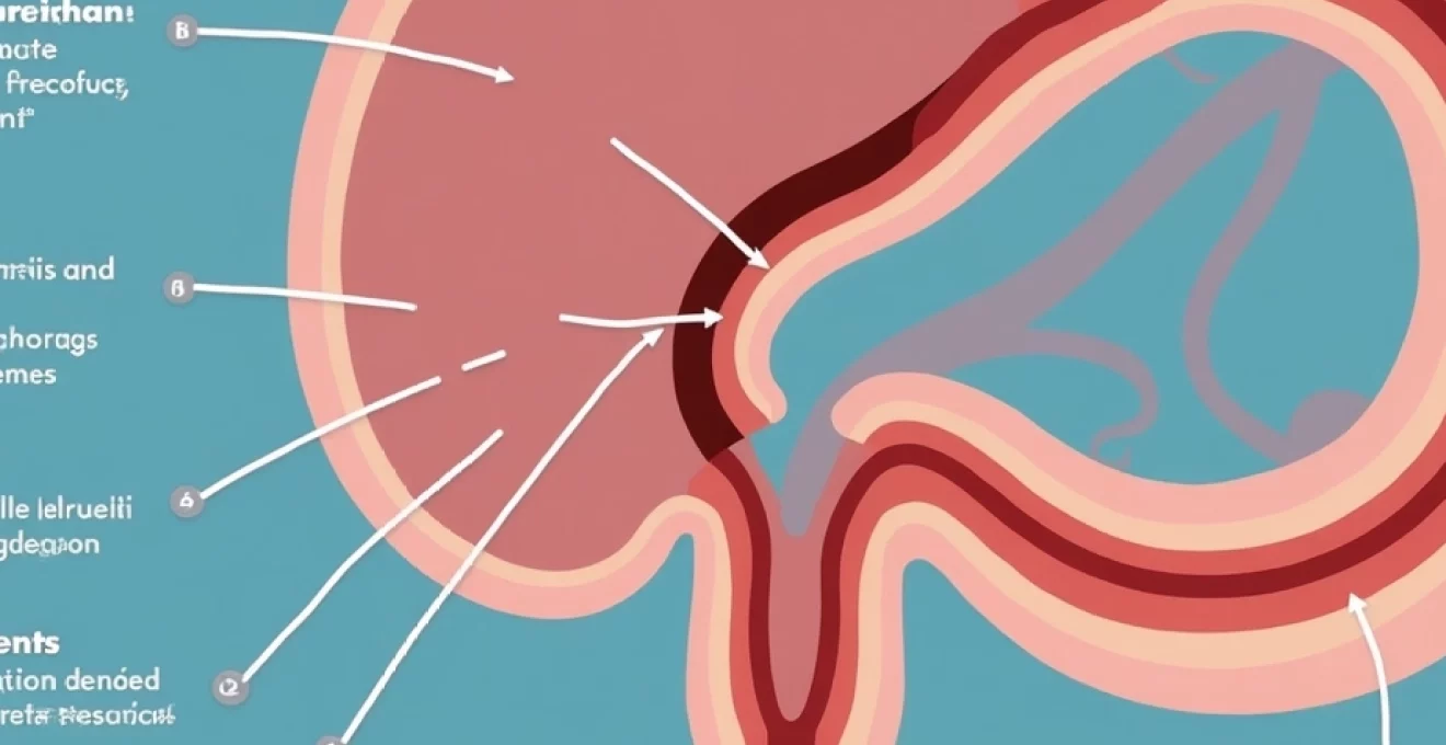
Understanding DJ stent anatomy and removal procedure
The double-J stent design represents a sophisticated solution to maintaining ureteral patency during healing or obstruction. These devices feature distinctive coiled ends that secure the stent within both the renal pelvis and bladder, preventing migration whilst facilitating continuous urine drainage. The central portion maintains ureteral dilation, ensuring adequate flow even in the presence of oedema or external compression.
Double-j stent design and materials: silicone vs polyurethane
Modern DJ stents utilise either silicone or polyurethane materials, each offering distinct advantages during placement and removal procedures. Silicone stents provide excellent biocompatibility and flexibility, reducing tissue irritation during dwelling periods. However, their softer composition can complicate removal in cases of significant encrustation or prolonged placement beyond recommended timeframes.
Polyurethane variants offer superior tensile strength and resistance to fragmentation during extraction procedures. This enhanced durability proves particularly valuable when removing stents that have remained in situ for extended periods, though they may generate increased irritative symptoms during the dwelling phase. The choice between materials often influences post-removal symptom severity and duration.
Cystoscopic removal technique using flexible and rigid cystoscopes
Cystoscopic stent removal employs either flexible or rigid instrumentation, with technique selection influencing patient comfort during and after the procedure. Flexible cystoscopy generally produces less urethral trauma and reduced post-procedural dysuria, though visualisation quality may be compromised in certain anatomical configurations. The gentler approach typically results in faster symptom resolution and improved patient tolerance.
Rigid cystoscopy offers superior optical clarity and instrument manoeuvrability, particularly beneficial when grasping encrusted or malpositioned stents. However, the increased urethral manipulation often correlates with more pronounced post-removal symptoms, including burning sensations and frequency that may persist for 48-72 hours. Understanding these technique-related differences helps set appropriate patient expectations.
Local anaesthesia vs sedation protocols during extraction
Anaesthetic protocols significantly impact both procedural comfort and post-removal symptom patterns. Topical lidocaine gel application provides adequate surface anaesthesia for routine removals, though deeper discomfort during stent manipulation remains poorly controlled. This limitation often results in involuntary muscle spasms and increased post-procedural inflammation.
Conscious sedation with midazolam and fentanyl offers superior patient comfort and procedural conditions, particularly for anxious individuals or complicated removals. However, sedated patients may experience delayed recognition of post-procedural symptoms and require extended monitoring periods. The choice between approaches should consider individual patient factors and procedural complexity.
Stent encrustation and biofilm formation impact on removal
Prolonged stent dwelling periods inevitably lead to mineral deposition and biofilm formation, significantly complicating removal procedures and intensifying post-extraction symptoms. Calcium phosphate and struvite crystals accumulate along the stent surface, creating rough textures that traumatise surrounding tissues during withdrawal. This mechanical irritation often produces more severe and prolonged discomfort compared to routine removals.
Biofilm development facilitates bacterial colonisation and chronic inflammation, contributing to persistent symptoms even after successful stent extraction. The inflammatory cascade triggered by biofilm disruption can perpetuate urinary frequency, dysuria, and flank discomfort for several days post-removal. Early stent removal within recommended timeframes significantly reduces these complications and associated patient morbidity.
Expected Post-Removal pain patterns and duration
Post-removal pain manifestations vary considerably amongst patients, influenced by factors including stent dwelling time, individual pain tolerance, and underlying urological conditions. Understanding typical symptom patterns enables appropriate patient counselling and reduces anxiety associated with normal recovery processes. Most patients experience some degree of discomfort, though symptom severity and duration remain highly variable.
Immediate Post-Procedural discomfort: first 24-48 hours
The initial 24-48 hour period following DJ stent removal typically presents the most intense symptoms, as inflamed tissues respond to the sudden removal of foreign material. Patients commonly report sharp, cramping sensations in the lower abdomen and pelvis, often described as similar to menstrual cramps or bladder spasms. These symptoms generally peak within 6-12 hours post-removal before gradually subsiding.
Pain intensity during this acute phase correlates with stent dwelling duration, with devices placed for seven days or less paradoxically causing more severe post-removal symptoms. This counterintuitive finding suggests that tissue adaptation occurs over time, with longer placement periods allowing better accommodation and reduced inflammatory response upon removal.
Dysuria and frequency symptoms: normal resolution timeline
Burning sensations during urination represent the most commonly reported post-removal symptom, affecting up to 80% of patients to varying degrees. This dysuria typically begins immediately following the procedure and gradually diminishes over 48-72 hours as urethral and bladder inflammation subsides. The intensity often correlates with the degree of procedural trauma and individual tissue sensitivity.
Urinary frequency accompanies dysuria in most patients, with normal voiding patterns typically resuming within 3-5 days post-removal. The bladder’s readjustment to the absence of foreign material requires time, as chronic irritation gradually resolves and normal detrusor function returns. Persistent frequency beyond one week warrants further evaluation for potential complications.
Flank pain characteristics following ureteral stent extraction
Flank discomfort following stent removal manifests differently from the colicky pain associated with acute obstruction, typically presenting as a dull ache or pressure sensation in the costovertebral angle region. This pain pattern reflects ureteral readjustment to normal peristaltic function after prolonged stenting. Most patients describe the sensation as mild to moderate, rarely requiring prescription analgesics for adequate control.
The duration of flank symptoms varies considerably, with most patients experiencing resolution within 24-48 hours. However, individuals with pre-existing hydronephrosis or ureteral strictures may experience prolonged discomfort as the collecting system readjusts to normal drainage patterns. Severe or worsening flank pain suggests potential complications requiring prompt urological evaluation.
Haematuria duration and associated discomfort levels
Microscopic haematuria following DJ stent removal occurs in virtually all patients, though visible blood in the urine affects approximately 60-70% of individuals. This bleeding typically presents as light pink discolouration that gradually clears over 2-3 days as traumatised tissues heal. The presence of small clots during the first 24 hours remains normal and rarely indicates significant complications.
Associated discomfort from haematuria usually remains mild, manifesting as slight burning or pressure sensations during voiding. Patients should expect gradual colour normalisation, though intermittent pink-tinged urine may persist for up to one week. Bright red bleeding or the passage of large clots warrants immediate medical evaluation to exclude significant injury or bleeding complications.
Differentiating normal recovery from complications
Distinguishing between expected post-removal symptoms and pathological complications requires careful attention to symptom patterns, timing, and severity. Normal recovery follows predictable patterns with gradual symptom improvement, whilst complications typically present with worsening symptoms, fever, or severe pain that fails to respond to conservative measures. Early recognition of concerning features enables prompt intervention and prevents serious morbidity.
Research demonstrates that patients with stents dwelling for more than seven days show significantly reduced post-removal pain, suggesting tissue adaptation over time reduces inflammatory responses upon extraction.
Urinary tract infection signs: temperature, WBC count, and nitrites
Urinary tract infections represent one of the most common complications following stent removal, occurring in approximately 5-15% of patients despite prophylactic measures. Early recognition relies on identifying the classic triad of fever, dysuria, and urinary frequency that persists or worsens beyond the expected 48-72 hour recovery period. Temperature elevation above 38.5°C accompanied by rigors suggests possible urosepsis requiring urgent intervention.
Laboratory markers including elevated white blood cell counts and positive urine nitrites provide objective confirmation of infection, though clinical symptoms often precede laboratory abnormalities. Patients should monitor for cloudy, malodorous urine accompanied by worsening pelvic pain or new-onset constitutional symptoms. The presence of pyuria exceeding expected post-procedural levels warrants prompt antibiotic therapy and culture-directed treatment.
Ureteral stricture development and associated pain patterns
Ureteral stricture formation represents a rare but serious complication that may manifest days to weeks following stent removal. Unlike normal post-removal discomfort, stricture-associated pain typically worsens progressively and may present with features of acute obstruction including severe flank pain, nausea, and vomiting. The pain often demonstrates a colicky nature, distinguishing it from the dull ache associated with normal recovery.
Early stricture development may present subtly with persistent low-grade flank discomfort and gradually worsening urinary symptoms. Patients may notice decreased urine output or changes in voiding patterns that fail to improve with time. Prompt recognition and intervention remain crucial to prevent irreversible renal damage and preserve long-term kidney function.
Retained stent fragments and persistent symptoms
Stent fragmentation during removal, whilst uncommon with modern materials, can result in persistent foreign body symptoms requiring additional intervention. Retained fragments typically cause ongoing irritative symptoms including frequency, dysuria, and intermittent haematuria that fail to resolve within expected timeframes. The symptoms often fluctuate in intensity but rarely completely resolve without fragment removal.
Imaging studies including plain radiography or CT scanning can identify retained fragments, particularly those containing radiopaque markers. Patients experiencing persistent symptoms beyond one week should undergo evaluation to exclude retained material, as fragments may serve as nidi for stone formation or chronic infection. Endoscopic retrieval usually provides definitive treatment with excellent outcomes.
Bladder perforation risk factors and warning signs
Bladder perforation during stent removal remains an exceedingly rare complication, typically associated with aggressive manipulation of severely encrusted devices or anatomical abnormalities. Warning signs include severe suprapubic pain, inability to void, and visible haematuria with clot passage. The development of peritoneal signs or abdominal distension suggests possible perforation with urinary extravasation.
Risk factors for perforation include prolonged stent dwelling time, significant encrustation, and previous bladder surgery or radiation exposure. Patients with these predisposing factors require careful monitoring during and after removal procedures. Early recognition and appropriate management, including catheter drainage and antibiotic therapy, typically result in favourable outcomes without long-term sequelae.
Pain management strategies after DJ stent removal
Effective post-removal pain management employs a multimodal approach combining pharmacological interventions with supportive care measures. Non-steroidal anti-inflammatory drugs (NSAIDs) provide excellent symptom control for most patients, addressing both pain and the underlying inflammatory process. Ibuprofen 400mg three times daily or diclofenac 50mg twice daily typically provides adequate analgesia whilst minimising adverse effects.
Alpha-blockers such as tamsulosin 0.4mg daily can significantly reduce bladder spasms and improve voiding comfort during the recovery period. These medications relax smooth muscle fibres in the bladder neck and urethra, facilitating easier urination and reducing associated discomfort. Treatment typically continues for 3-5 days until symptoms resolve naturally.
Non-pharmacological interventions play important supportive roles in symptom management. Increased fluid intake promotes healing through dilution of irritating substances and mechanical flushing of the urinary tract. Warm baths or heat application to the lower abdomen can provide significant comfort for cramping sensations. Patients should aim for 2-3 litres of fluid daily unless contraindicated by cardiac or renal conditions.
Studies indicate that female patients demonstrate a 2.41-fold increased risk of experiencing post-removal pain, emphasising the importance of gender-specific counselling and pain management strategies.
When to seek urgent medical attention
Certain symptoms following DJ stent removal warrant immediate medical evaluation, as they may indicate serious complications requiring urgent intervention. High fever exceeding 38.5°C, particularly when accompanied by rigors or confusion, suggests possible urosepsis and demands emergency treatment. Similarly, complete inability to urinate or severe reduction in urine output may indicate acute obstruction or bladder dysfunction.
Severe, uncontrolled pain that fails to respond to prescribed analgesics requires prompt assessment to exclude complications such as perforation or retained fragments. The development of new neurological symptoms, including leg weakness or sensory changes, though rare, may suggest epidural complications requiring immediate neurosurgical consultation. Patients experiencing chest pain, difficulty breathing, or signs of pulmonary embolism should seek emergency care immediately.
Persistent heavy bleeding with passage of large clots or inability to void due to clot retention necessitates urgent urological intervention. The development of abdominal distension, particularly when accompanied by severe pain and vomiting, may indicate bowel complications or severe infection requiring immediate surgical evaluation. Early recognition and prompt treatment of these complications significantly improve patient outcomes and prevent long-term morbidity.
Recovery timeline and Follow-Up protocols
The typical recovery timeline following DJ stent removal follows predictable patterns, with most patients experiencing gradual symptom improvement over 5-7 days. Initial discomfort usually peaks within 12-24 hours post-removal before steadily declining. Complete resolution of irritative symptoms typically occurs within one week, though some patients may experience intermittent mild symptoms for up to two weeks.
Follow-up protocols vary depending on the underlying condition necessitating stent placement and individual patient factors. Routine post-removal appointments typically occur 1-2 weeks after the procedure, allowing assessment of symptom resolution and identification of potential complications. Patients with complex histories or high-risk features may require more frequent monitoring, including laboratory studies and imaging as clinically indicated.
Long-term follow-up requirements depend on the underlying pathology and treatment outcomes. Patients with recurrent stone disease require ongoing surveillance and preventive measures, whilst those with benign strictures may need periodic imaging to assess for recurrence. The development of chronic symptoms or recurrent infections warrants comprehensive evaluation to identify treatable causes and optimise long-term outcomes. Understanding these timelines helps patients maintain realistic expectations and seek appropriate care when symptoms deviate from normal recovery patterns.